Search
Close this search box.

MIMA2

Microscopy and Imaging of Microorganisms, Animal Models and Food

MIMA2 Core Facility, at the forefront of imaging technologies, supports diverse biological investigations. It addresses scientific projects, from medical imaging in animal models to microbial ecosystems’ study. Innovating through partnerships, MIMA2 bridges academia and industry, enhancing regional, national, and European scientific ecosystems while maintaining rigorous quality standards.

Vlad Costache

Platform Operational manager

Romain Briandet

Scientific manager

anonymous user

Anne Couturier-Tarrade

Scientific manager

MIMA2 Core Facility focuses on phenotyping of microorganisms, embryos, organoids and live large animal model systems, within its four thematic poles. MIMA2-Micalis pole (MIMO) is specialized in confocal and electron microscopy of class 2 pathogens and commensals such as viruses, bacteriophages, bacteria, and fungi.

Taking advantage of its optical and electronic microscopes, MIMA2 Core Facility demonstrates expertise in biofilm high content screening, ultrastructural analysis of cells, tissue, extracellular vesicles (EVs) and provides proficiency in organ clearing techniques (lightsheet imaging of large samples) and environmental scanning electron microscopy analysis.

The objectives of MIMA2 are to provide access to cutting-edge imaging techniques and to offer comprehensive scientific and technical assistance to users for their research endeavors.

Practical information

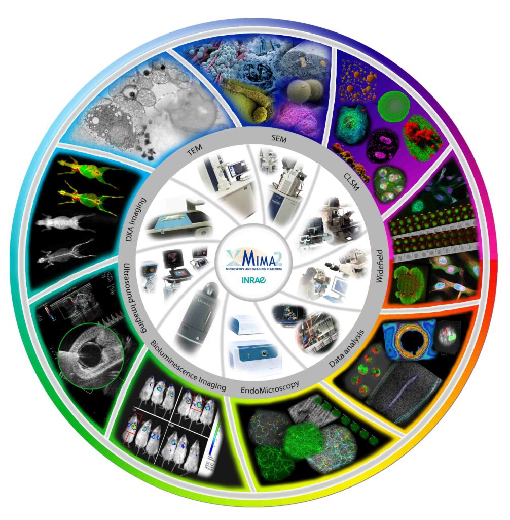
The MIMA2 Core Facility combines complementary equipment giving the possibility to perform imaging work from the nanoscopic scale to the whole animal, using live cells/pathogens or previously processed samples, as well as model and even domestic animals.

The platform is localized on the campus of the Jouy-en-Josas INRAE near Paris. It is open to external users, from academia and private sector.

Practical know-how and laboratory space are available for users, including veterinary and surgical competence, sample processing protocols and automated processing equipment. Some of the work (mainly microscopic observations) can also be performed in confined L2 environment. Platform users can work in full autonomy after being trained and provided legal requirements in the case of animal experimentation. Users can ask for the platform to perform the analyses.

The microscopic and live imaging equipment of the platform are supported by four INRAE research units in Jouy-en-Josas INRAE campus (UMR 1198 BREED, UMR 1319 MICALIS, UMR 1313 GABI, UMR 0892 VIM) and are altogether referenced as Outil Collectif N°35 and labeled regionally as a strategic platform ISC (Infrastructure Scientifique Collective). MIMA2 is also identified in the network of Paris-Saclay’s university department of life sciences within the innovation domain “life quality, health and food”.

The MIMA2 platform offers key resources in light and electron microscopy, as well as in medical imaging.

Team members

Vlad COSTACHE

Zoé COMMANDER

Romain BRIANDET

Julien DESCHAMPS

Microscopy of Embryos and Organoids (Pole IMEO – Buildings 440 & 230):

  • Pierre ADENOT – Engineer (BREED)
  • Matthieu SIMION – Engineer (BREED)


Medical Imaging (Pole CIMA – Building 402):

  • Christophe RICHARD – Engineer (BREED)
  • Valérie GELIN – Technician (BREED)
  • Gwendoline MORIN – Technician (BREED & SAAJ)


Cell Ultrastructure (Pole UC – Building 440):

  • Christine LONGIN – Engineer (GABI)
  • Martine LETHEULE – Engineer (BREED & GABI)
  • Thierry MEYLHEUC – Engineer
  • Alexis CANETTE – Engineer 

Our quality pledges

Logo LRQA
LRQA Certified ISO 9001:2015 – 00040132 for Transmission Electron Microscopy Activities