Search
Close this search box.

ProCeD

Prokaryotic Cell Development

We are interested in understanding how dynamic molecular interactions are regulated in time and in space to form functional machineries that establish long-range orders and cellular functions in bacteria, with a particular focus on the bacterial cytoskeleton and on the cell envelope, at the frontline of environmental and host-pathogen interactions.

To this end, we combine cutting-edge high-resolution fluorescence microscopy and spectroscopy techniques with powerful genetic, biophysical, biochemical and systems biology approaches to determine mechanistic details underlying cellular and developmental functions in bacteria. We study in particular (but not only) the rod-shaped Gram-positive model bacterium Bacillus subtilis and the Gram-negative ESKAPE pathogen Pseudomonas aeruginosa (classed by the World Health Organization (WHO) as priority 1 (critical) pathogen for R&D in antimicrobial research). Our current work follows 3 main research axes:

Research axis

GFP-MreB imaged by  SIM-TIR

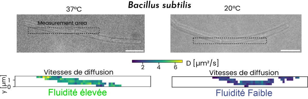
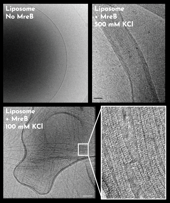
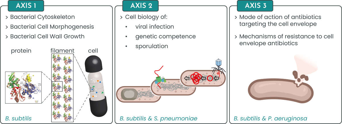
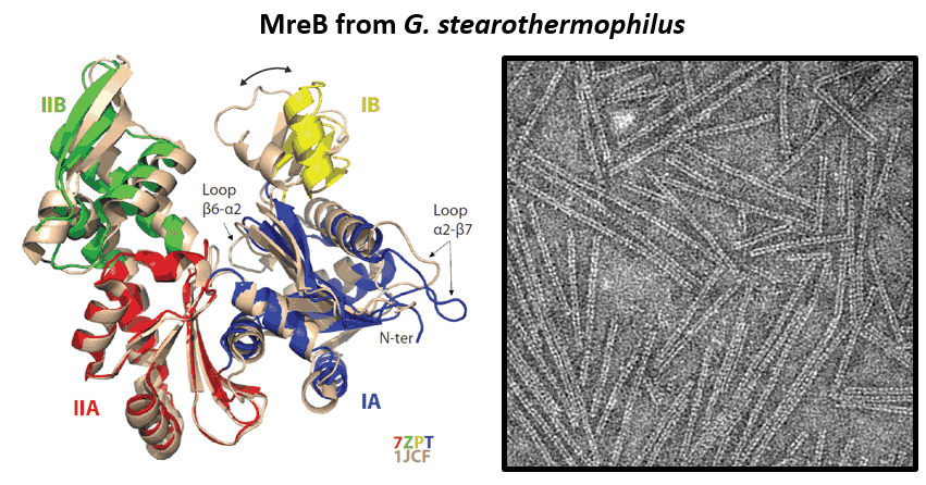
Little is known about the mechanisms determining cell shape, and the larger questions concerning morphogenesis are the same in prokaryotic and eukaryotic systems. How is structural information acquired and maintained? How is cell shape spatially and temporally regulated? In bacteria, the extracellular cell wall (a micrometer-scale 3D polymer network and the most prominent target for antibiotics) and the intracellular actin-like (MreB) cytoskeleton are major determinants of cell shape. We and others previously showed that MreB proteins assemble into membrane-associated nanofilaments that move processively around the cell periphery and control shape by organizing and orienting transmembrane enzymes that effect sidewall elongation in the extracellular space. The properties of MreB assemblies, the mechanistic details underlying their morphogenetic function and the interplay between MreB, the plasma membrane and the cell wall synthesizing machineries remain however to be elucidated.

MreB from G.stearothermophilus

In the core project of our lab, currently funded by an ERC Consolidator Grant, and previously by an ERC Starting Grant, we investigate the mechanistic details underlying cell wall growth, membrane organization and MreB function(s) using a combination of in vivo and in vitro approaches. Research on the bacterial actins is important because some mechanisms might be conserved in higher organisms and/or provide evolutionary cues. Conversely, research on the bacteria-specific cell wall is also highly important because it provides potential strategies for the development of antimicrobials at a time when multiple antibiotics resistance has become a major health concern. Our long-term goals are to understand general principles of bacterial cell morphogenesis and bacterial actins to provide mechanistic templates and new reporters for the screening of novel antibiotics.

Prokaryotic Cell Development
S. pneumoniae

We are also exploring the cell biology of cellular processes occurring across the bacterial cell wall and/or requiring cell wall remodeling. Currently, these include phage infection, genetic competence and sporulation. These are in general collaborative projects mainly funded by the ANR (French National Research Agency).

  • Together with the lab of Paulo Tavares (i2BC, Gif-sur-Yvette, France), specialized in phage biology, we study the cell biology of phage infection. The biological system under study is the well-characterized bacteriophage SPP1 that infects B. subtilis. We discovered that during infection, SPP1 builds spatially independent factories for viral genome replication and assembly of viral particles in the bacterial cytoplasm. Our collaboration aims to characterize molecular and cellular mechanisms underlying the formation of these factories.
  • In collaboration with the team of Nathalie Campo (LMGM, Toulouse,France) and Christophe Grangeasse (MMBS, Lyon), we are studying competence for genetic transformation of the human pathogen Streptococcus pneumoniae (the streptococcus). Our project focuses on the molecular characterization of a programmed cell division arrest that occurs during competence, with the aim of understanding how the pneumococcus coordinates competence for genetic transformation with its life cycle.
  • When facing severe nutritional stress, B. subtilis forms highly resistant endospores. The pathway to sporulation is probably the best-studied developmental program in bacteria. However, the function of many genes expressed during this process remains a mystery. In collaboration with Ciaran Condon (IPBC, Paris,France) and Adriano Henriques (ITQB, Oeiras (or Lisbon), Portugal), we are trying to understand the role of an orphan ribonuclease that is expressed specifically in the mother cell during sporulation and may have a role in spore coat assembly.

We investigate the mode of action of antibiotics targeting the cell envelope in both B. subtilis and the ESKAPE, WHO priority 1 pathogen P. aeruginosa.

  • In B. subtilis, we are performing integrated, system biology studies of the mode of action of antibiotics targeting different steps of cell wall (peptidoglycan) synthesis that we then couple with mechanistic studies (Axis 1).
  • In P. aeruginosa, we are studying the mode of action and resensitization to the last resort antibiotic colistin as well as the mechanisms controlling P. aeruginosa infection modes. P. aeruginosa is able to switch from a planktonic to a sedentary biofilm lifestyle corresponding to acute and chronic infections, respectively. In parallel to the cell biology studies, we are implementing high resolution fluorescence imaging tools and methods to image live P. aeruginosa cells at different scales: single cell – biofilm – human airway epithelial cells, to mimic the lung environment. Our Pseudomonas research benefits of excellent collaborations with the teams of Steve Porter (Exeter University, UK), Romain Briandet (Micalis, B3D), Craig MacLean (Oxford University, UK), Florence Vincent (AFMB, Aix-Marseille University, France), Rosa del Campo (University Hospital Ramon y Cajal of Madrid, Spain), Vinciane Saint-Criq (Micalis, ProbiHôte) and Stefano Pagliara (Exeter University, UK).

Team members

Claire-Jing ROUCHET

Ingrid ADRIAANS

Cyrille BILLAUDEAU

Arnaud CHASTANET

Rut CARBALLIDO LOPEZ

Armand LABLAINE

Merve Nur TUNÇ

Aurélien BARBOTIN

Dimitri JUILLOT

Matteo GERARD

  • Charlène CORNILLEAU (2013-2023, Engineer / PhD / Post-doc)
  • Clara LOUCHE (2023-2023, Engineer)
  • Sana AFENSISS (2022-2023, Engineer)
  • Laurie-Anne TRIVAL (2021-2023, Engineer)
  • Caroline PERON-CANE (2019-2023, Post-doc)
  • Sarah BENLAMARA (2020-2022, Engineer)
  • Maria-Victoria PREJEAN (2010-2022, Technician)
  • Ipek ALTINOGLU (2019-2021, Post-doc)
  • Vlad COSTACHE (2020-2021, Post-doc)
  • Yunjiao LU (2017-2020, PhD)
  • Magali VENTROUX (2015-2020, Engineer)
  • Peggy MERVELET (1998-2020, Technician)
  • Alexis CARREAUX (2019-2020, Engineer)
  • Wei MAO (2017-2020, Post-doc)
  • Ruth KEARY (2014-2019, Post-doc)
  • Sriram TIRUVADI-KRISHNAN (2017-2019, Post-doc)
  • Elisa BRAMBILLA (2016-2017, Post-doc)
  • Benoît TESSON (2014-2017, Post-doc)
  • Alba DE SAN EUSTAQUIO CAMPILLO (2013-2017, PhD)
  • Xavier HENRY (2014-2016, Post-doc)
  • Alex DAJKOVIC (2012-2016, Post-doc)
  • Pierre FLORES (2012-2016, PhD)
  • Cécile FERRET (2013-2016, Engineer)
  • Nicolas MIROUZE (2012-2016, Post-doc)
  • Smita CHAUHAN (2014-2016, Engineer)
  • Céline MONNIOT (2014-2015, Post-doc)
  • Zhizhong YAO (2013-2015, Post-doc)
  • Calum MACKICHAN (2008-2013, PhD)
  • Yoan AH-SENG (2011-2012, Post-doc)
  • Célia PALCY (2010-2012, Engineer)
  • Anne-Stefanie RUEFF (2008-2011, PhD)
  • Elodie MARCHADIER (2005-2009, PhD)
  • 2024: Philéas LARCHER, Hind OUKLFIF, Sumudu Upatissa
  • 2023: Thomas HAINAUT, Joseph NAKFOUR, Emma WALKER
  • 2022: Ali DAKROUB, Louise DESTOUCHES, Lilia HUYNH, Elena THOMANN
  • 2021: Lea WAGNER
  • 2017: Lorenzo CARRE, Marine GUITON
  • 2015: Sina KROKOWSKI, Quentin SANDRO
  • 2014: Andreia TAVARES
  • 2010: Anaiss SAQUET

Key points

News